Figure 1.
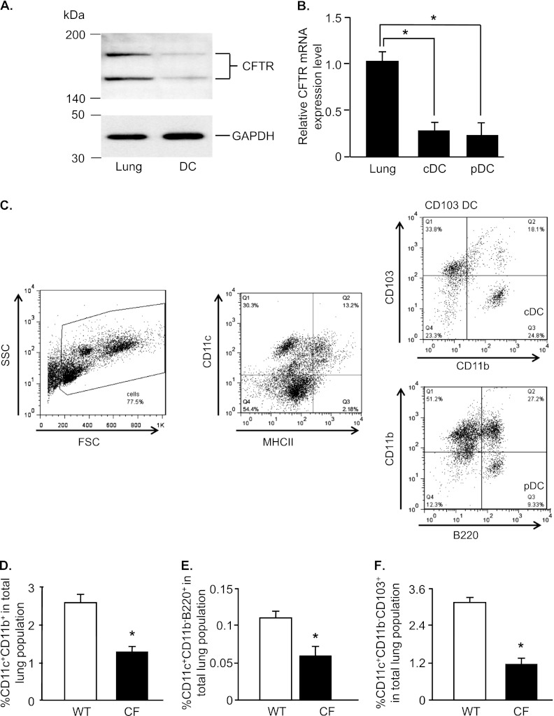
Cystic fibrosis transmembrane regulator (CFTR) expression in dendritic cells (DCs), and decreased percentage of DCs in CFTR knockout (CF) mice. DCs (CD11c+) were isolated from the lungs of wild-type (WT) C57BL/6 mice by magnetic beads, and were further separated into conventional DCs (cDCs; CD11c+CD11b+) or plasmacytoid DCs (pDCs; CD11c+CD11b−B220+) by fluorescence-activated cell sorting. (A) Western blot analysis of CFTR protein expression in DCs and all lung cells. (B) CFTR mRNA expression in cDCs, pDCs, and all lung cells by real-time RT-PCR, normalized to the expression of glyceraldehyde 3–phosphate dehydrogenase mRNA. Data are presented as the means ± SEMs of n = 5 mice/group for one of two independent experiments. (C) Diagram of gating strategy of cDCs and pDCs by flow cytometry. The percentages of cDCs (D), pDCs (E), and CD103-positive (CD103+) DCs (F) from CF mice and WT control mice were quantified by flow cytometry. Data are expressed as the means ± SEMs of the percentages of stained cells of all lung cells. Data shown are representative of six independent experiments, each with n = 4–5 mice/group. *P < 0.05. SSC, side scatter; FSC, forward scatter; MHCII, major histocompatibility complex class II molecules; Q, quadrant.

