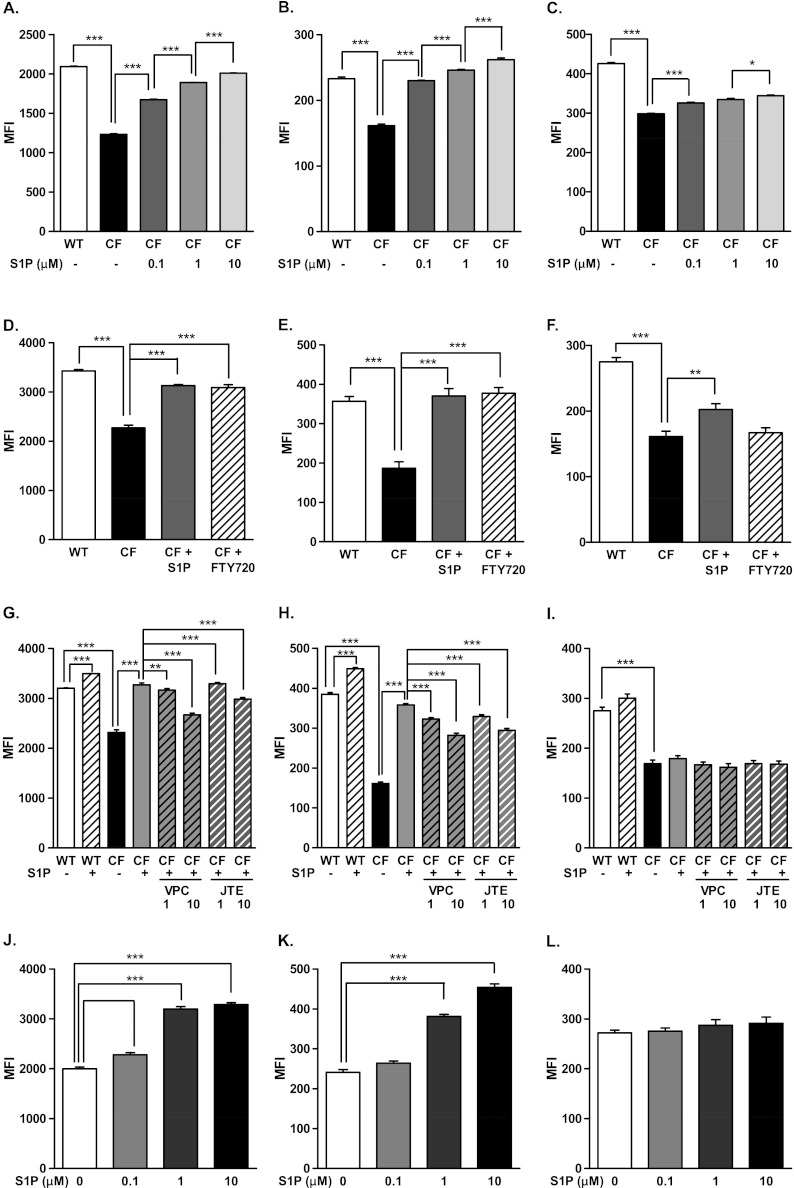
Figure 7.
Effects of S1P on maturation and activation of DCs cultured in vitro in CF BALF. Lung DCs from WT mice were cultured in CF BALF supplemented with 0.1, 1.0, and 10.0 μM S1P (A–C), or 20 nM of the S1P analogue FTY720 or 1 μM S1P (D–F), or 1 and 10 nM of the S1P receptor inhibitors JTE013 (JTE) or VPC23019 (VPC) (G–I) for 24 hours. CF lung DCs were cultured in CF BALF supplemented with 0.1, 1.0, and 10.0 μM S1P (J–L) for 24 hours. The surface expression of MHCII (left panels), CD40 (middle panels), and CD86 (right panels) was assessed by flow cytometry. Data represent the means ± SEMs of mean fluorescence intensity. *P < 0.05. **P < 0.01. ***P < 0.001.

