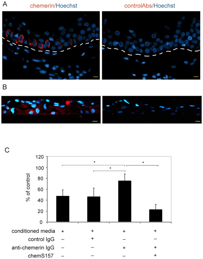
Figure 1. Keratinocyte-derived chemerin displays anti-bacterial activity.
Paraffin sections of normal, shoulder skin biopsies (A) or chest keratinocytes grown in 3D culture for 1 week (B) were stained for chemerin or control rabbit Abs (red), with Hoechst counterstain to detect cell nuclei (blue). The slides were examined by fluoresce microscopy. Dotted lines in A indicate location of epidermis. Scale bar = 10 µm. Data are representative of three different donors. The antimicrobial activity of conditioned media from 3D cultures of keratinocytes (conditioned media) was tested against E. coli using the microtitre broth dilution assay (C). Where indicated, the conditioned media were first treated with sepharose-conjugated anti-chemerin Ab (anti-chemerin IgG), sepharose-conjugated control IgG (control IgG), or anti-chemerin Ab followed by recombinant chemerinS157 (chemS157) at 20 ng/ml. The results are expressed as the mean ± SD of four independent experiments. Statistically significant differences are indicated by asterisks (p≤0.01, Student's t test).

