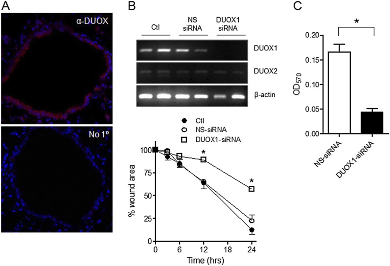
Figure 2.
Wound responses in MTE cells are mediated by dual oxidase–1 (DUOX1). (A) Immunofluorescence analysis of DUOX protein in lung tissue from C57BL6 mice. Top: Immunofluorescence detection of α-DUOX (red) and nuclear counterstaining with 4,6-diamidino-2-phenylindole (DAPI) (blue). Bottom: Similar analysis without primary (1°) antibody. (B) DUOX1 expression in MTE was silenced by small interfering (si)RNA, as confirmed by RT-PCR (above). The effects of DUOX1 silencing on wound closure were evaluated in a scratch wound assay (below). Mean values ± SEs of 4–6 replicates from 2–3 experiments are presented. *P < 0.05, compared with nontarget (NS)–siRNA. (C) MTE cells transfected with DUOX1 siRNA or NS-siRNA were seeded on fibronectin-coated inserts for analyses of cell migration, as measured by the absorbance (optical density; OD) at 570 nm of crystal violet–stained migrated cells (n = 5). *P < 0.05, compared with NS-siRNA.

