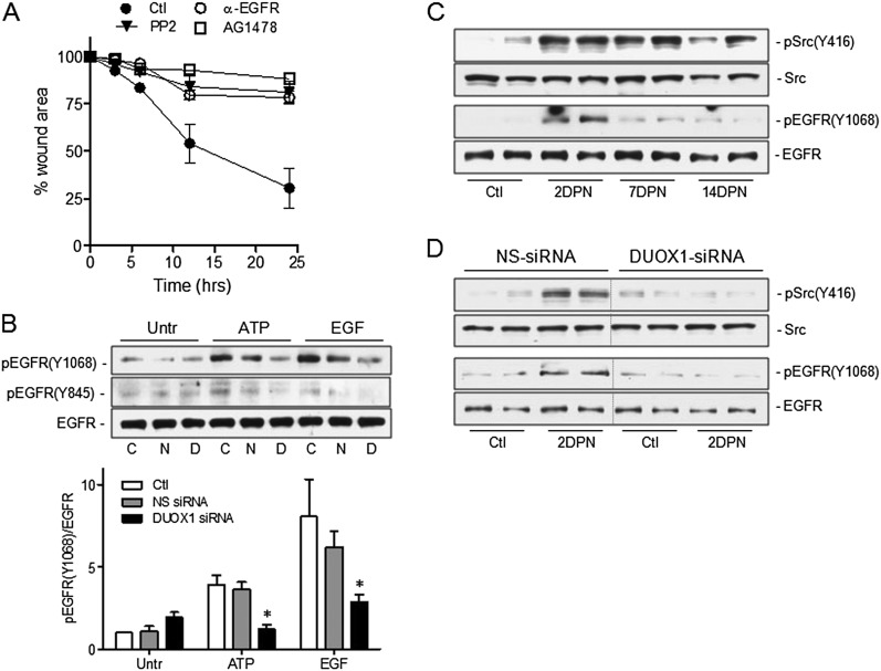
Figure 5.
Role of Src–epidermal growth factor receptor (EGFR) signaling in DUOX1-mediated epithelial wound responses. (A) Analysis of wound closure rates in scratched MTE monolayers in the absence or presence of the Src inhibitor PP2 (10 μM), the EGFR tyrosine kinase inhibitor AG1478 (10 μM), or a blocking EGFR monoclonal antibody (4 ng/ml). Data represent the mean ± SE (n = 3–5). (B) Western blot analysis of pEGFR (Y1068) and total EGFR in untreated MTE cells (Untr) or MTE cells transfected with DUOX1 siRNA or NS-siRNA, after 10-minute stimulation with exogenous ATP (100 μM) or epidermal growth factor (EGF; 10 ng/ml). Representative Western blots and relative band intensities from three separate experiments are presented. C, control; N, NS-siRNA; D, DUOX1 siRNA. *P < 0.05, compared with corresponding control samples. (C) Analysis of phosphorylated forms of EGFR and Src (pEGFR Y1068 and pSrc Y416) in lung-tissue homogenates of mice collected 0, 2, 7, or 14 days after an intraperitoneal injection of naphthalene. (D) Similar analysis of pEGFR (Y1068) or pSrc (Y416) in lung tissues obtained 2 days after naphthalene injection (2 DPN) or vehicle control (Ctl) and instillation of DUOX1 siRNA or NS-siRNA. Representative blots show results from two mice.

