Figure 6.
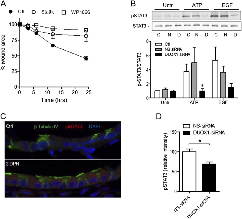
DUOX1-dependent activation of signal transducer and activator of transcription–3 (STAT3) phosphorylation in epithelial wound responses. (A) Analysis of wound closure rates in scratched MTE monolayers in the absence or presence of the two structurally unrelated inhibitors of STAT3, Stattic (10 μM), or WP1006 (5 μM). Data represent the mean ± SE (n = 3–5). (B) Western blot analysis of pSTAT (Y705) and total STAT3 in MTE cells after transfection with DUOX1 siRNA or NS-siRNA, and stimulation with ATP (100 μM) or EGF (10 ng/ml) for 10 minutes. Representative Western blots and relative band intensities from three separate experiments are shown. C, control; N, NS-siRNA; D, DUOX1 siRNA. *P < 0.05, compared with corresponding control samples. (C) Immunofluorescence imaging of pSTAT3 in lung-tissue sections obtained 2 days after vehicle (Ctl) or naphthalene injection (2DPN). (D) Quantification of pSTAT3 immunofluorescence intensity within the epithelium was performed on 2-day naphthalene-exposed mice receiving NS siRNA or DUOX1 siRNA, using Metamorph software. Data are based on average intensities, determined in at least three airways per mouse, and mean values ± SEs from five animals are shown. *P < 0.05, compared with NS-siRNA.

