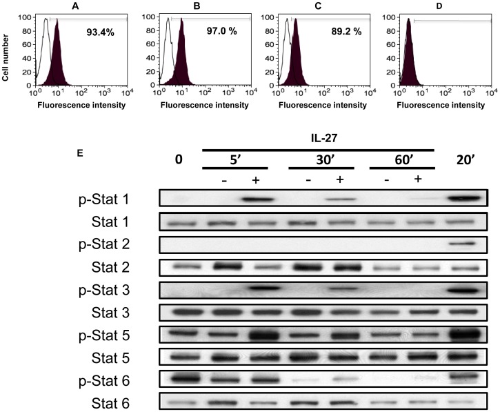
Figure 1. Expression levels of IL-27Rα (WSX-1) in DCs and STAT activation following IL-27 treatment.
A–D: The expression level of the IL-27 receptor (WSX-1) was measured using flow cytometry on the following cells: A, Freshly isolated monocytes B, iDCs C, mDCs D, LLC. These cells were stained with either a PE-conjugated anti-WSX-1 (black) antibody or an isotype control (white). Data indicates a representative result from a single donor from three independent experiments. The numbers in the figures show percentage of positive cells, and the Mean Fluorescence Intensity (MFI) of monocytes, iDCs and mDCs were 8.75, 9.13 and 6.3, respectively. E: Western blotting was used to measure the phosphorylation status of STAT-1, -2, -3, -5, and -6 in IL-27 or untreated iDCs at a number of time points. For each phosphorylated STAT measured, the unphosphorylated protein was also measured to show that the profiles were not due to loading differences.

