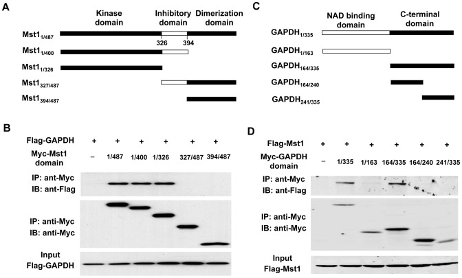
Figure 3. Identification of Mst1 and GAPDH interaction sites.
A, schematic representation of Mst1 deletion mutants. B, flag-GAPDH expression vector in combination of either empty vector or expression vectors of Myc-Mst1 mutants were cotransfected into HEK293T cells. Extracted proteins were precipitated by anti-Myc antibody and then separated by 12% SDS-PAGE. The transferred membrane was immunoblotted with either HRP-conjugated anti-FLAG or HRP-conjugated anti-Myc antibody. Lysates were also immunoblotted with anti-Flag antibody to show the expression levels of Flag-GAPDH in HEK293T cells. C, schematic representation of GAPDH deletion mutants. D, Flag-Mst1 expression vector in combination of either empty vector or expression vectors of Myc-GAPDH mutants were cotransfected into HEK293T cells. Extracted proteins were precipitated by anti-Myc antibody and then separated by 12% SDS-PAGE. The transferred membrane was immunoblotted with either HRP-conjugated anti-FLAG or HRP-conjugated anti-Myc antibody. Lysates were also immunoblotted with anti-Flag antibody to show the expression of Flag-Mst1 in HEK293 cells.

