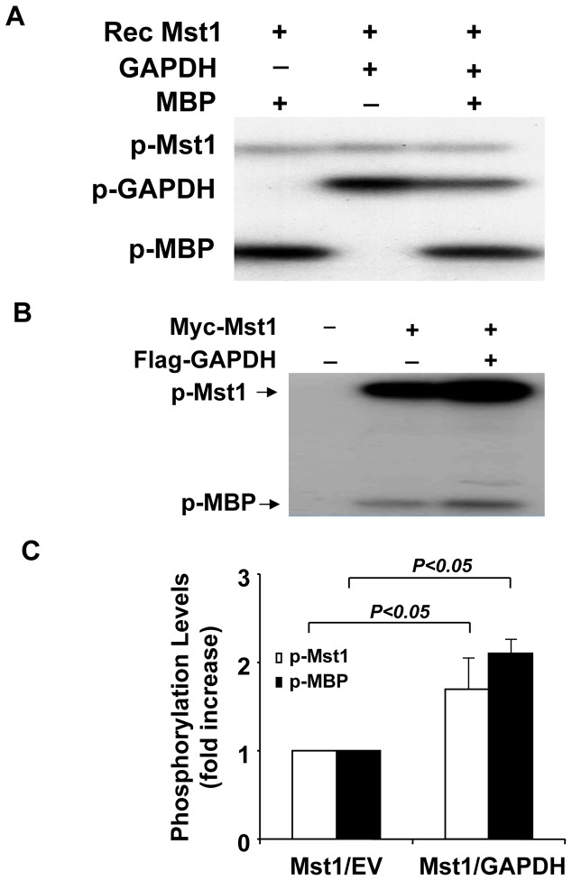
Figure 5. Activation of Mst1 kinase by GAPDH.
A, 0.1 µg active Mst1 was incubated with either 4 µg of recombinant GAPDH or MBP for 60 min in the presence of 32P-ATP in an in vitro phosphorylation assay. Phosphorylation was detected by autoradiography. B, Mst1 immunoprecipitated from HEK293 cells transfected with either Myc-Mst1 or Myc-Mst1 plus Flag-GAPDH expression vectors was incubated with 2 µg MBP for the in vitro kinase assay. Kinase assay was carried out in the presence of 32P-ATP for 60 min. Phosphorylation was detected by autoradiography. C, Phosphorylation levels of Mst1 and MBP were quantified by densitometry of autoradiograms. Values are means ± SEM obtained from 3 experiments.

