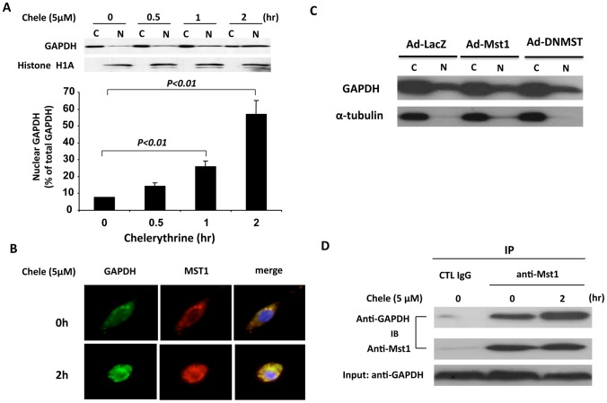
Figure 6. Nuclear translocation of GAPDH and Mst1 during cardiomyocyte apoptosis.
A, NRVMs were treated with chelerythrine (5 µM) for different time points as indicated. Cytoplasmic and nuclear fractions were isolated and then subjected to western blot analysis using ant-GAPDH and anti-histone H1A antibodies. The distribution of GAPDH in the cytoplasmic and nuclear fractions was analyzed by densitometric analysis. Values are means ± SEM obtained from 4 experiments. B, Unstimulated NRVMs or NRVMs stimulated with chelerythrine (5 µM) for 2 hours were fixed and stained with anti-GAPDH monoclonal antibody and rabbit polyclonal anti-Mst1 antibody and processed for confocal imaging. The merged images show clear colocalization of these 2 proteins in cytoplasm in ustimulated cells and translocation and colocalization of these 2 proteins in nucleus in response to chelerythrine. C, NRVMs were transduced with either Ad-LacZ or Ad-Mst1 or Ad-DNMST (MOI = 30). 48 hr after transduction, cells were treated with chelerythrine (5 µM) for 1 hour. Cytoplasmic and nuclear fractions were isolated and then subjected to western blot analysis using anti-GAPDH and anti--tubulin antibodies. D, Unstimulated NRVMs or NRVMs stimulated with chelerythrine (5 µM) for 2 hours were lysed and then subjected to immunoprecipitation with either normal IgG or anti-Mst1 antibody. Immunocomplexes were then separated by 15% SDS-PAGE and transferred membrane was immunoblotted with either anti-GAPDH or Mst1 antibody.

