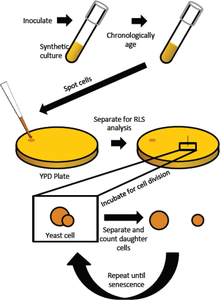
Figure 1. Diagram depicting the chronological lifespan to replicative lifespan assay.
Cells are aged chronologically in synthetic complete (SC) medium with either 2% glucose (control) or 0.05% glucose (DR). At different age points, an aliquot is removed from the chronologically aging culture and spotted onto rich medium (YPD) for replicative lifespan analysis. Individual cells are arrayed on the YPD plate and those replicative lifespan is determined.

