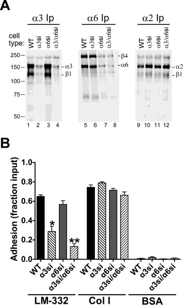
Fig. 1.
Specific silencing of α3 and α6 integrin subunits in GS698.Li prostate carcinoma cells. a Parental (WT), α3-silenced (α3si), α6-silenced (α6si), and doubly silenced cells (α3/α6si) were surface-labeled with biotin and extracted with 1% Triton X-100. Integrins α3β1, α6β4, or α2β1 were immunoprecipitated (Ip) from normalized lysates, and then analyzed by blotting with IRdye 800-streptavidin. b WT, α3si, α6si, and α3/α6si cells were allowed to adhere to wells coated with laminin-332 (LM-332), collagen I (Col I), or BSA for 20 min in serum-free medium. Non-adherent cells were removed by rinsing, and adherent cells were fixed and quantified by staining with crystal violet. Results are presented as a fraction of total cells input, as measured in poly-L-lysine control wells. Two independent trials that gave similar results were pooled (for total of 8 wells per cell type/condition) Error bars indicate S.E.M. *Significantly less than WT parental cells, ANOVA with Tukey-Kramer t test, (p<0.001); **Significantly less than WT (p<0.001) and significantly less than α3si (p<0.05), ANOVA with Tukey-Kramer t test.

