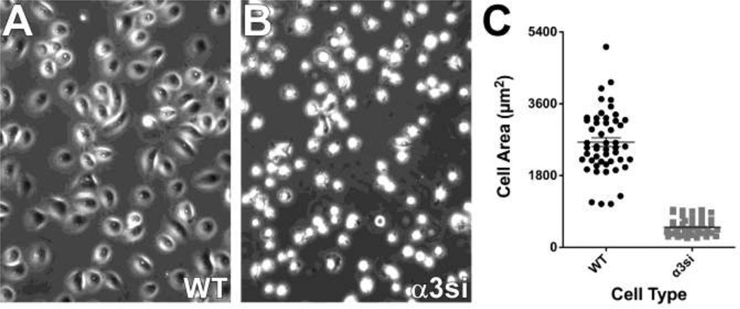
Fig. 3.
Severely impaired spreading of α3 silenced cells on LM-332. a & b WT and α3si cells were plated in serum-free medium on LM-332-coated glass bottom culture dishes. After 30 min for cell attachment and spreading, cells were photographed with a 20X objective. c 50 cells of each type were quantified using ImageJ to measure cell area. The α3si cells were significantly less well spread, p < 0.0001, unpaired t test.

