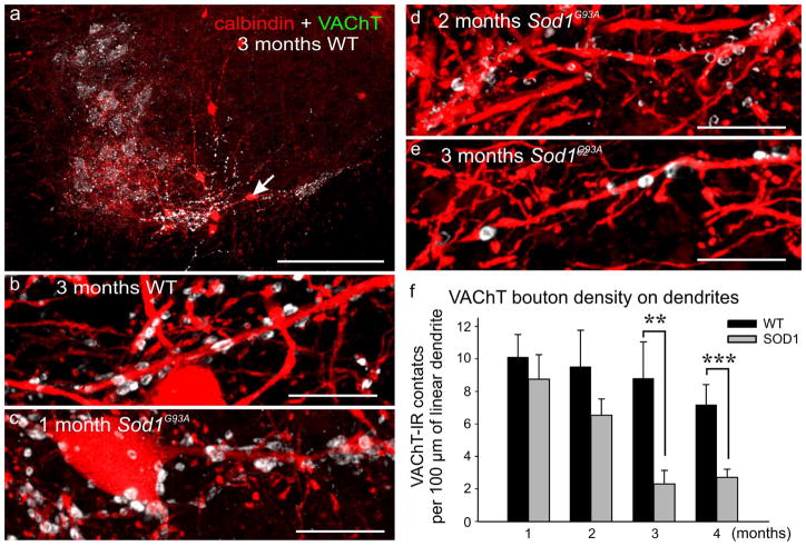
Figure 4.
VAChT-IR synapses on Renshaw cells are profoundly altered in Sod1G93A mice, starting at 2 months of age. (a) Low and (b) high magnification confocal images of VAChT immunoreactivity (green) and Renshaw cells (red) in WT animals. (c-e) Images of calbindin-IR Renshaw cell dendritesin 1, 2 and 3 month old Sod1G93A mice. VAChT-IR fills most boutons in contact with WT Renshaw cells VAChT-IR boutons of 2 month Sod1G93A animals appear hollow and the immunoreactivity concentrates at the periphery of the bouton (e). Few boutons remain in 3 month Sod1G93A mice. (f) Quantification of VAChT-IR contacts on Renshaw cell dendrites. No significant differences in contact density were found in WTs of different ages (p=0.653, One-Way ANOVA). Contact density was significantly reduced in 3 month (p<0.01, Bonferroni corrected t-test) and 4 month (p<0.001, Bonferroni corrected t-test)old Sod1G93A animals compared to their age-matched controls, but not in 1 and 2 month Sod1G93A animals. Error bars indicate SEMs. Scale bars: in a, 250 μm; in b-e, 10 μm.(A version of this figure in which white-green has been changed for magenta-green is supplied as supplementary figure 3).

