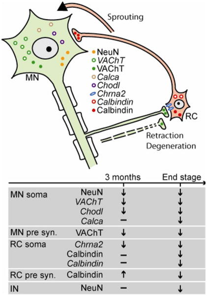
Figure 7.
Schematic drawing summarizing the changes in motor neuron and Renshaw cells during disease progression. In a motor neuron cell body, NeuN and VAChT together with the fast motor neuron marker Chodl decrease first and Calca decreases later. In the receiving Renshaw cell body, Chrna2 decreases before a decrease in Calbindin can be detected. Simultaneously with these changes in gene expression motor axon synapses on Renshaw cells become abnormal and dennervate Renshaw cells dendrites, while Renshaw cell synapses seem to undergo compensatory innervation of remaining NeuN-IR motor neurons.

