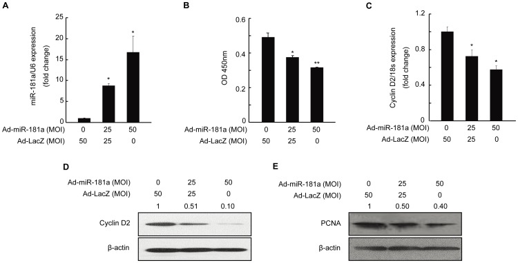
Figure 2. Inhibition of proliferation of and relevant gene expression in mouse granulosa cells (mGC) by miR-181a.
mGC was infected with Ad-miR-181a (multiplicity of infection, MOI = 0, 25, and 50) for 48 h. (A) MiR-181a level was measured by qRT-PCR. (B) Result of a CCK-8 assay examining the proliferation of mGC. (C) qRT-PCR and (D) Western blot analysis of cyclin D2 mRNA and protein levels, respectively, in mGC. (E) Protein level of proliferating cell nuclear antigen (PCNA) as determined by Western blotting. Relative protein levels were measured by densitometry using Quantity One Software and normalized to β-actin, Ad-LacZ group; the ratios were presented above the Western blot bands. *p<0.05, **p<0.01, compared with Ad-LacZ group.

