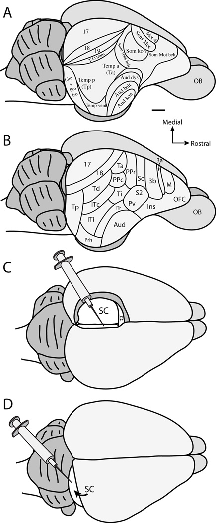
Figure 1.
Organization scheme of tree shrew cortex based on A. Casseday et al., 1979, and B. adapted from Wong and Kaas (2009). C. Illustration of anatomical tracer placement after aspiration of the contralateral hemisphere and retraction of the ipsilateral hemisphere to the injected superior colliculus. D. Illustration of anatomical tracer placement after retraction of the occipital lobe to visualize the caudal aspect of the superior colliculus. See Table 2 for abbreviations. Scale bar is 2mm.

