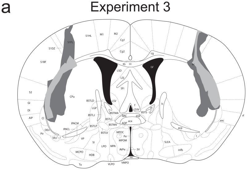
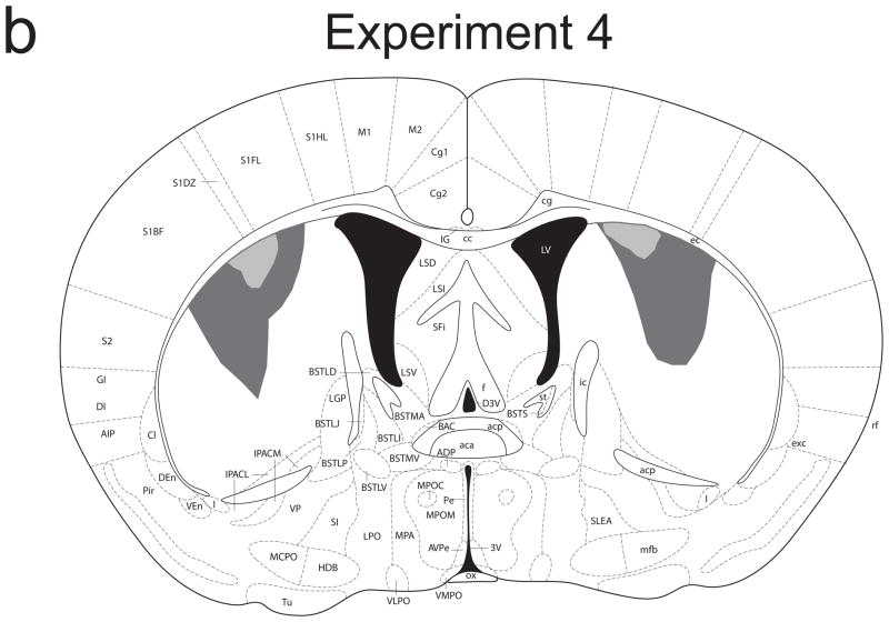
Figure 4.
Schematic representation of NMDA-induced excitotoxic lesions of the lateral (a) and intermediate (b) dorsal striatum in Experiments 3 and 4, respectively. Shaded areas represent the minimum (light gray) and maximum (dark gray) extent of the lesions for mice included in all analyses (adapted from Paxinos & Franklin, 2001).


