
Figure 3.
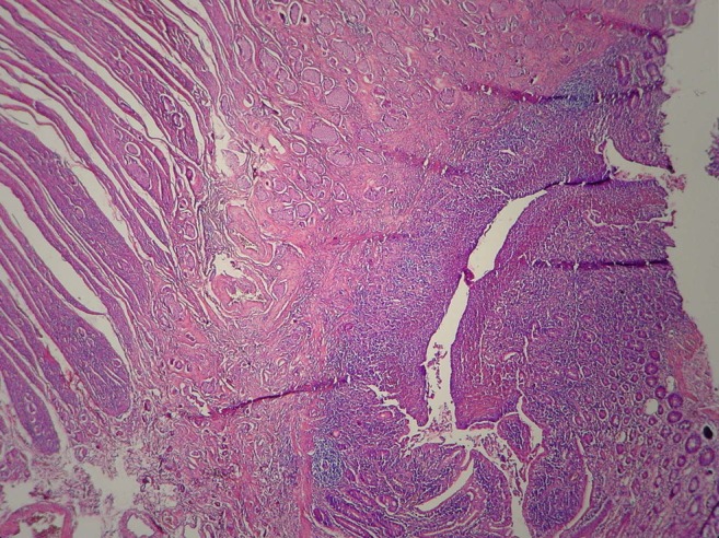
Microscopy sample showing the mucosa and submucosa of ileum invaded by several solid nests of cells, corresponding to type I growth and well-differentiated neuroendocrine tumour (G1).
Official websites use .gov
A
.gov website belongs to an official
government organization in the United States.
Secure .gov websites use HTTPS
A lock (
) or https:// means you've safely
connected to the .gov website. Share sensitive
information only on official, secure websites.
Microscopy sample showing the mucosa and submucosa of ileum invaded by several solid nests of cells, corresponding to type I growth and well-differentiated neuroendocrine tumour (G1).