Fig. 4.

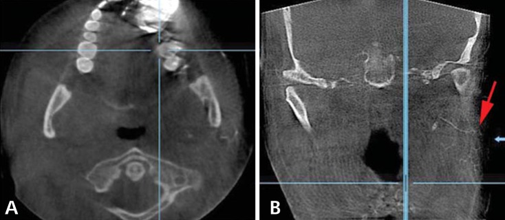
Axial cut (A) and coronal cut (B) of CBCT sialography show stenosis of the ductules with punctuated appearance of the gland (arrow).

Axial cut (A) and coronal cut (B) of CBCT sialography show stenosis of the ductules with punctuated appearance of the gland (arrow).