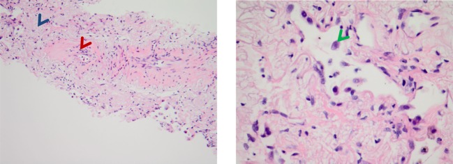
Figure 3.

Case 1 pathology—stereotactic body radiation therapy site: (left) H&E stain showing hyalinisation of vessels (red arrow) and reactive interstitial fibrosis (blue arrow); (right) H&E stain showing peribronchial reactive atypical pneumocytes (green arrow).
