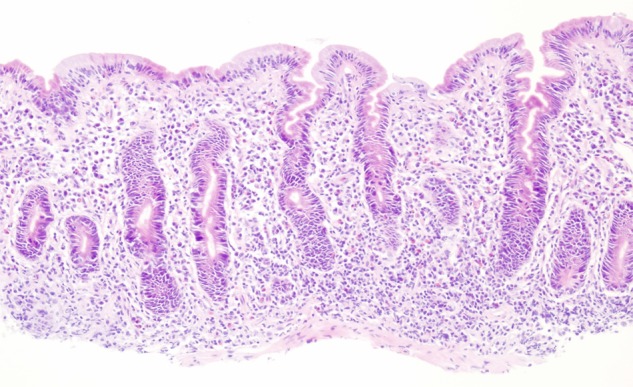
Figure 3.

Duodenal biopsy. Histology shows subtotal to total villous atrophy, increased cellularity in the lamina propria due to lymphoplasmacellular infiltrates, increased number of intraepithelial lymphocytes, loss of goblet cells, Paneth cells and endocrine cells. An increased number of mitotic figures and apoptotic bodies at the crypt base were noted.
