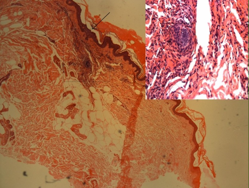
Figure 1.

Photomicrograph of skin biopsy showing vasculitis in the superficial dermal vessels with fibrinoid necrosis of vessel wall and neutrophilic infiltration, consistent with leucocytoclastic vasculitis at a magnification of ×25, H&E stain. Area denoted in the box shown on the right-hand corner at higher magnification.
