
Figure 3.
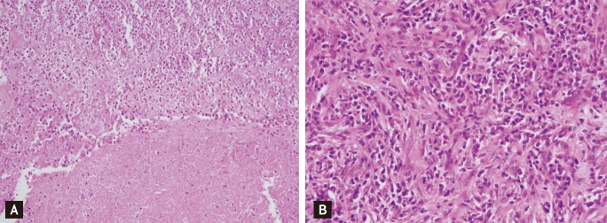
Histopathological examination shows extensive central necrosis with abscess formation (A, H&E, ×200). In the periphery, massive plasma cells and scattered lymphocytes with fibrosis were seen (B, H&E, ×400).
Official websites use .gov
A
.gov website belongs to an official
government organization in the United States.
Secure .gov websites use HTTPS
A lock (
) or https:// means you've safely
connected to the .gov website. Share sensitive
information only on official, secure websites.
Histopathological examination shows extensive central necrosis with abscess formation (A, H&E, ×200). In the periphery, massive plasma cells and scattered lymphocytes with fibrosis were seen (B, H&E, ×400).