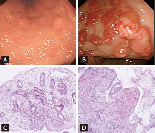
Figure 1.
Endoscopic and histological findings of gastric polyposis associated with portal hypertension. (A) Initial esophagogastroduodenoscopy shows several raised erosions in the lower body. (B) Multiple polyps with superficial erosions or erythema are noted 15 months later. (C) Pathologic specimen shows foveolar hyperplasia, edematous lamina propria, and ectatic capillaries (H&E, × 100). (D) There is erosion of, and marked granulation tissue in, the lamina propria C D (H&E, × 100).

