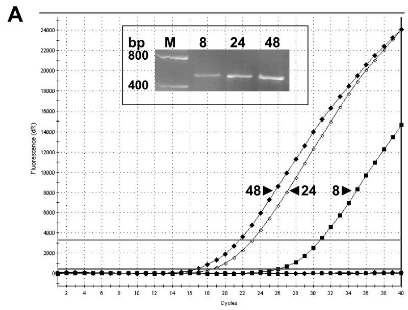
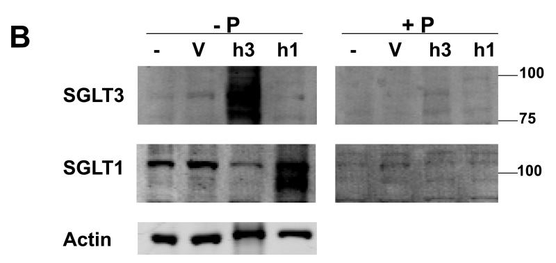
Fig. 1. Human SGLT1 and SGLT3 specific antibodies.
COS-7 cells transiently transfected with vectors for over-expression of hSGLT1 (h1) or hSGLT3 (h3) were used to examine the reactivity and specificity of antibodies. (A) Total RNA samples were prepared 8, 24, and 48 hours post transfection and used in RT-real time PCR with hSGLT3-specific primers. Amplification profiles are shown in the graph and gel electrophoresis analysis of PCR products are shown in the inset. M, DNA size marker; bp, base pairs. (B) Forty eight hours post transfection with h1 or h3 vectors, total cell lysates were prepared from COS-7 cells and used in Western blot. Cell lysates from transfection vehicle (V) and untreated (−) cells were used as controls. Blots were probed with 1 μg/ml of hSGLT1 or hSGLT3 antibody and infrared (IR) signals were detected at 800 nm; -P, antibody alone. Above experiments were repeated with peptide-blocked antibodies; +P. Blots were re-probed with actin antibody and IR detection was carried out at 700 nm. Positions of MW size markers are shown.


