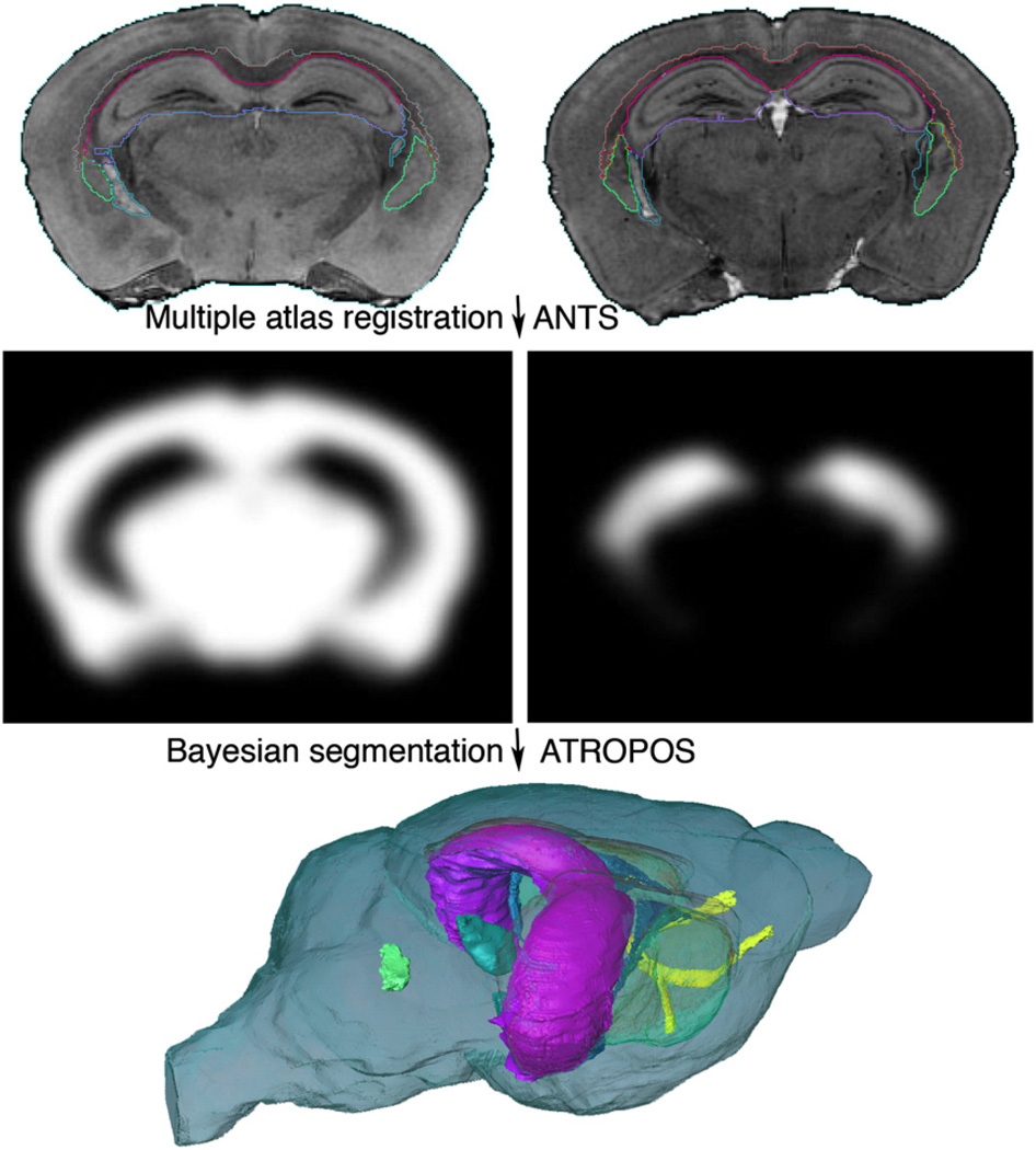
Fig. 7.
Multiple specimen atlases were used to generate probabilistic priors. The location, intensity, and label priors can be used in a Bayesian segmentation framework to improve upon the accuracy of single atlas based segmentation. Five specimens were used to generate priors using ANTs; subsequently used for MRF segmentation using ATROPOS. The accuracy of multi-atlas segmentation was evaluated for 6 labeled regions including the anterior commissure (yellow), corpus callosum (orange), hippocampus (magenta), striatum (transparent green), substantia nigra (cyan), and ventricles (LV: blue; V4: solid green).

