Figure 1.
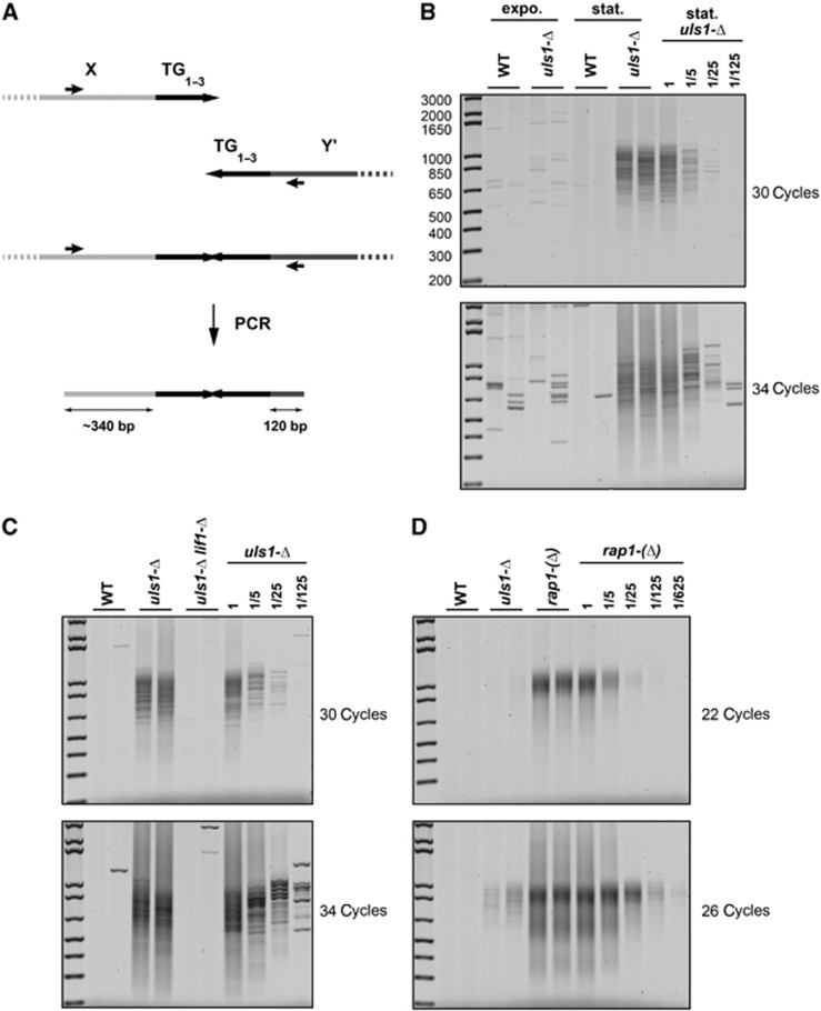
Loss of Uls1 causes telomere fusions by NHEJ. (A) Schematic representation of the relative positions of the primers used for PCR amplification. The average wild-type telomere length is ∼300 bp. (B) Telomere fusions accumulate in stationary uls1-Δ cells. Two independent cultures of strains Lev346 (WT) and RL71 (uls1-Δ) were grown exponentially in rich medium (expo.) and allowed to reach stationary phase in 6 days (stat). Fusions between X and Y′ telomeres were amplified by PCR with 30 and 34 cycles. Genomic DNA from RL71 (uls1-Δ) cells in stationary phase was diluted serially to provide a semi-quantitative estimation of the method sensitivity. (C) Telomere fusions in uls1-Δ cells are NHEJ dependent. Strains 212-12a and 213-4a (WT), 213-7b and 213-9c (uls1-Δ) and 195-28a (uls1-Δ lif1-Δ) were grown to stationary phase. Telomere fusions were amplified with 30 and 34 cycles. Genomic DNA from 213-7b (uls1-Δ) cells was diluted serially. (D) Telomere fusions in uls1-Δ are less frequent than in rap1-(Δ) cells. Strains 210-3d and 211-1a (WT), 211-8d and 211-10b (uls1-Δ) and 209-1c and 209-2b (rap1-(Δ)) were grown to stationary phase. Telomere fusions were amplified with 22 and 26 cycles. Genomic DNA from 209-1c (rap1-(Δ)) cells was diluted serially to provide a semi-quantitative estimation of the relative difference of fusion frequencies.

