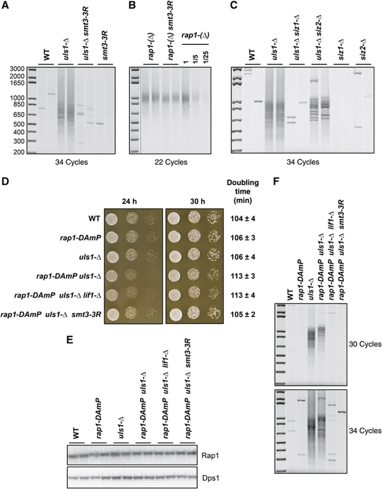
Figure 3.
Suppression of uls1-Δ by smt3-3R. (A) Telomere fusions are undetectable in usl1-Δ smt3-3R cells. Two independent cultures of strains RL179 (WT), RL183 (uls1-Δ), RL185 (uls1-Δ smt3-3R) and RL181 (smt3-3R) were grown to stationary phase. Telomere fusions were amplified with 34 cycles. (B) Telomere fusions remain frequent in rap1-(Δ) smt3-3R cells. Strains 169-1c and 169-15c (rap1-(Δ)) and 169-6b and 169-11a (rap1-(Δ) smt3-3R) were grown to stationary phase. Telomere fusions were amplified with 22 cycles. Genomic DNA from 169-1c rap1-(Δ) cells was diluted serially to provide a semi-quantitative estimation of the method sensitivity. (C) Uls1 suppression by Siz loss. Two independent cultures of strains 168-3d (WT) and 168-3d (uls1-Δ) and strains RL266-1 and RL266-2 (uls1-Δ siz1-Δ), RL267-1 and RL267-2 (uls1-Δ siz2-Δ), RL268-1 and RL268-2 (siz1-Δ) and RL269-1 and RL269-2 (siz2-Δ) were grown to stationary phase. Telomere fusions were amplified with 34 cycles. (D) Slow growth of rap1-DAmP uls1-Δ cells is suppressed by smt3-3R. Freshly growing cells of strains 195-19d (WT), 195-3a (rap1-DAmP), 195-7b (uls1-Δ), 195-8a (rap1-DAmP uls1-Δ), 195-6b (rap1-DAmP uls1-Δ lif1-Δ) and 195-1d (rap1-DAmP uls1-Δ smt3-3R) were serially diluted by 10-fold in water and spotted on a rich medium plate. Pictures were taken after 24 and 30 h at 30°C. Doubling times are from exponential growth in liquid-rich medium at 30°C. (Data shown are the mean and standard deviation of five independent samples). (E) Cultures from the same strains were grown exponentially and total urea-extracted proteins were analysed by western blotting with polyclonal antibodies directed against Rap1 (upper panel) and against RNA synthetase Dps1 (an internal control, lower panel). (F) Cultures from the same strains were grown to stationary phase and telomere fusions were amplified with 30 and 34 cycles.

