Figure 1.
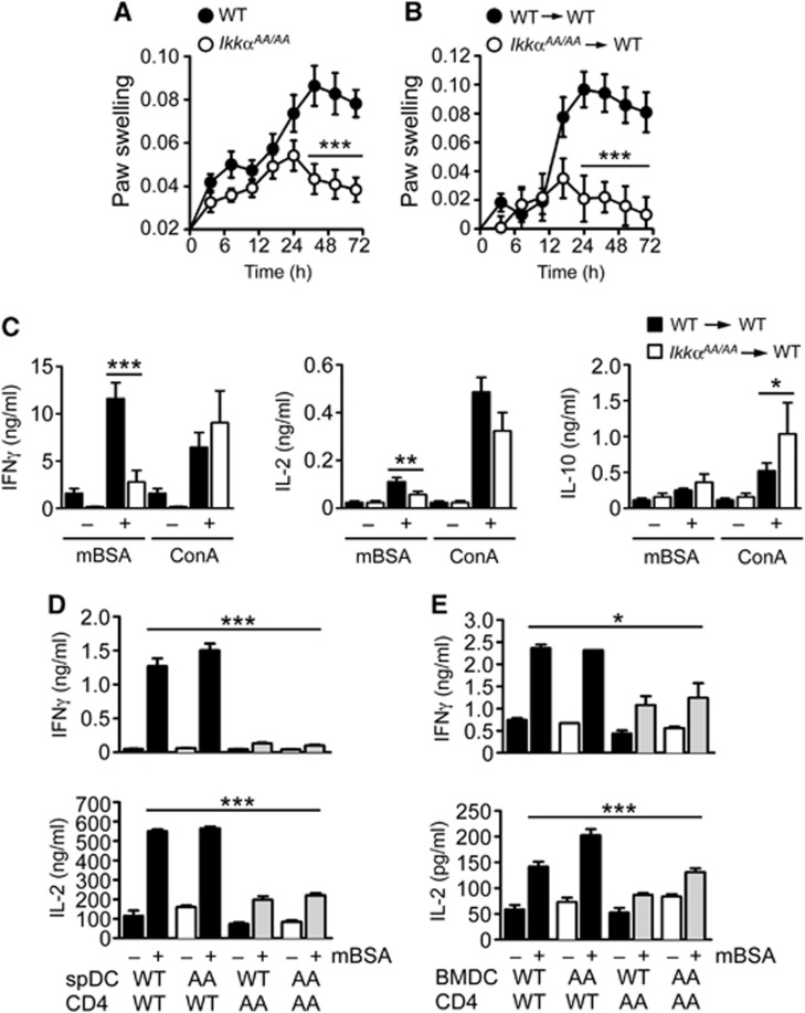
IKKα is required for CD4+ T-cell priming in vivo. (A) IkkαAA/AA and littermate control mice (WT), and radiation chimeras generated with WT and IkkαAA/AA bone marrow cells (B), were immunized intradermally (i.d.) with CFA/mBSA. Fourteen days after immunization, mice were injected with mBSA in the left footpad, PBS was injected into the right footpad as a control. Paw swelling was measured at the indicated time points by plethysmography as an index of antigen (mBSA)-induced inflammation. Data are represented as mean±s.e.m. of n=10, statistical analysis was performed with Mann–Whitney test; ***P≤0.001. (C) Splenocytes from CFA/mBSA-immunized WT and IkkαAA/AA chimeric mice were collected after 14 days and re-stimulated in vitro with mBSA or concanavalin A (ConA); IFNγ, IL-2 and IL-10 were measured in culture supernatants by ELISA after 72 h. Data are represented as mean±s.e.m. of n=10, statistical analysis was performed with Mann–Whitney test; *P≤0.05, **P≤0.005, ***P≤0.001. (D) CD4+ T cells were isolated from lymph nodes (LN) and spleen of CFA/mBSA-immunized WT and IkkαAA/AA (AA) mice after 14 days; T cells were co-cultured with CD11c+ DC isolated from spleens of naïve WT and IkkαAA/AA mice (spDC; D), or bone marrow-derived DC (BMDC; E), in the presence or absence of mBSA. IFNγ and IL-2 were measured in culture supernatants by ELISA after 72 h. Data are represented as mean±s.e.m. of n=6, statistical analysis was performed with Mann–Whitney test; *P≤0.05, ***P≤0.001.

