Figure 3.
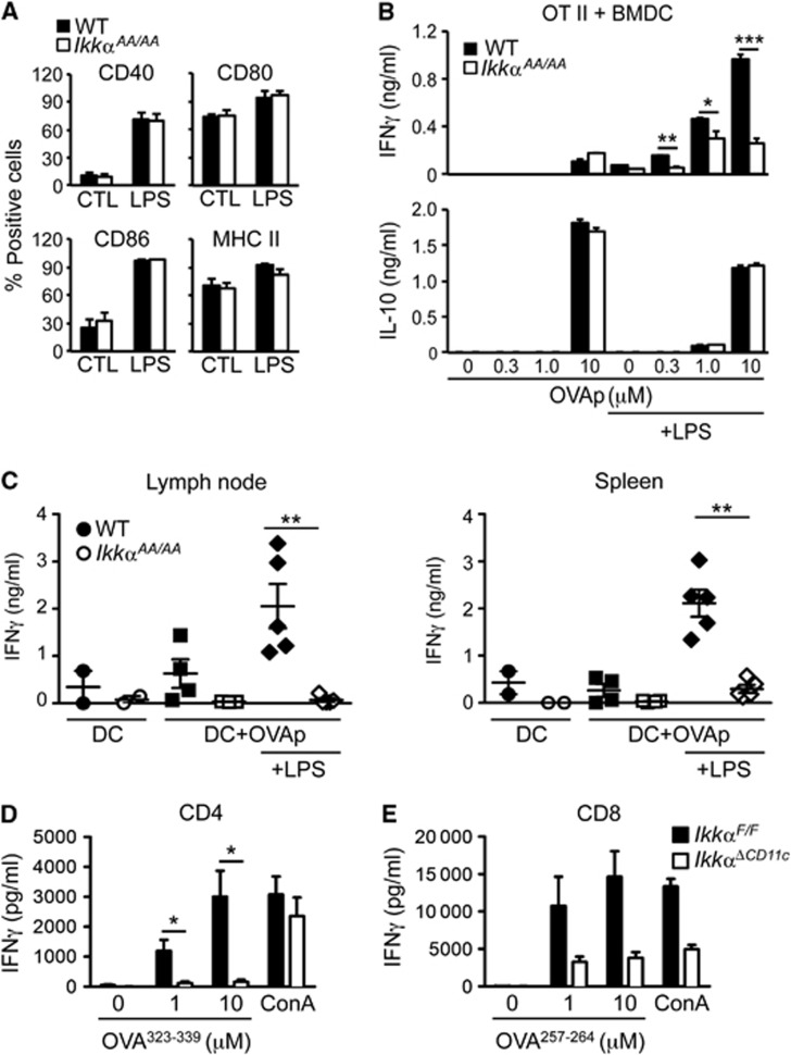
IKKα is required for TLR-induced functional maturation of DC. (A) FACS analysis of BMDC from WT and IkkαAA/AA mice after LPS (100 ng/ml)-induced maturation for 24 h. Data are represented as mean±s.e.m. of n=3. (B) BMDC from WT and IkkαAA/AA mice were loaded with MHC II-restricted OVA peptide (OVA323–339; OVAp) with and without LPS stimulation for 24 h before co-culture with naïve CD4+ OT-II T cells. IFNγ and IL-10 were measured in culture supernatants after 72 h. Data are represented as mean±s.e.m. of n=4, statistical analysis was performed with Student’s t-test; *P≤0.05, **P≤0.005, ***P≤0.001. (C) BMDC from WT and IkkαAA/AA mice were loaded with OVAp in the presence and absence of LPS for 24 h, 2.5 × 10⁵ DC were injected into the footpad of naïve WT mice. Seven days after DC injection, the popliteal LN and spleen were collected and single-cell suspensions prepared for antigen (OVAp) re-call assay in vitro; measured by IFNγ production in culture supernatants after 72 h. Data are represented as mean±s.e.m. of n=5, statistical analysis was performed with Mann–Whitney test; **P<0.005. (D, E) 106 OVA-specific CD4+ (OT-II; D) or CD8+ (OT-I; E) T cells were adoptively transferred to IkkαΔCD11c and IkkαF/F mice, before immunization with CFA/OVA i.d. The inguinal LN was collected after 5 days and single-cell suspensions prepared for antigen re-call assays with MHC II (OVA323–339) and MHC I (OVA257–264) specific peptides; IFNγ production was measured in culture supernatants after 72 h. Data are represented as mean±s.e.m. of n=3–6, statistical analysis was performed with Mann–Whitney test; *P<0.05.

