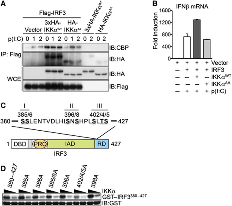
Figure 7.
IKKα regulates IRF3-medaited transcription downstream of IKKε/TBK1-mediated IRF3 phosphorylation. (A) HEK293-TLR3 cells were transfected with cDNA vectors expressing Flag-IRF3, HA-IKKαWT and HA-IKKαAA, in the presence of absence of p(I:C) as indicated. Protein extracts were prepared and immunoprecipitation (IP) of IRF3 was performed with anti-Flag antibody, co-IP of HA-IKKα and endogenous CBP was detected by IB analysis. Expression of Flag-IRF3, HA-IKKαWT and HA-IKKαAA was measured in total protein extracts (WCE) by IB. (B) In parallel experiments, RNA was extracted at 4 h and IFNβ expression measured by qRT–PCR. Representative data from at least two independent experiments are shown; qRT–PCR data are presented as mean±s.e.m. of three replicates. (C) Schematic representation of the seven potential phosphorylation sites in the C-terminal regulatory domain of IRF3 organized into three clusters: I—Ser385/Ser386; II—Ser396/Ser398; III—Ser402/Thr404/Ser405. DBD, DNA-binding domain; PRO, proline-rich domain; IAD, auto-inhibitory domain; RD, regulatory domain. (D) GST-IRF3380–427 and various mutant peptides were expressed and purified from bacteria and incubated with recombinant active IKKα in the presence of γ-32P-ATP, IKKα-mediated phosphorylation of GST-IRF3 peptides was quantified by autoradiography. IB analysis of GST was used a loading control for IRF3 substrate. Representative data from two independent experiments are shown.
Source data for this figure is available on the online supplementary information page.

