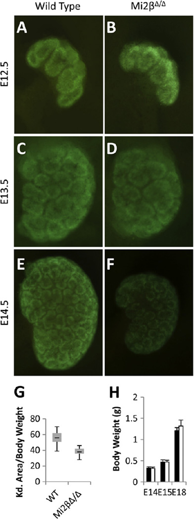
Fig. 2.
Deletion of Mi2β from cap mesenchyme cells results in renal hypoplasia. (A–F) Whole kidney images of GFP expressed from the Cre:GFP fusion protein under control of the Six2 BAC transgene in wild type and Mi2βΔ/Δ embryos at E12.5 (A, B), E13.5 (C, D), and E14.5 (E, F). Both wild type and Mi2βΔ/Δ kidneys were similar in size at E12.5 and E13.5 (A–D). Hypoplasia and reduction of GFP expression was evident at E14.5 in Mi2βΔ/Δ kidneys compared to wild type littermates (E, F). (G) Quantification of hypoplasia by relating kidney area to body weight of the embryo over a developmental stage period of E13.5 to E18.5. Deletion of Mi2β resulted in renal hypoplasia in 90% of mutants with an average 33% reduction in the area/body weight ratio. (H) Average body weights of wild-type and Mi2βΔ/Δ embryos. Body weights of wild type (black bars) and Mi2βΔ/Δ mutant (white bars) embryos were indistinguishable throughout development.

