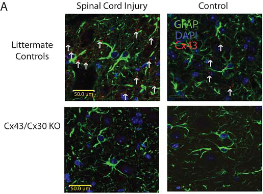
Fig. 1. Immunohistochemical staining of injured and noninjured spinal cord.
(A) Immunohistochemical staining of GFAP (green), DAPI (blue), and Cx43 (red) in longitudinal spinal cord slices of Cx43/Cx30 KO mice and littermate controls. As expected, in Cx43/Cx30 KO, GFAP-expressing cells express no Cx43. Meanwhile, littermate controls express Cx43 in GFAP-expressing cells and exhibit an upregulation of CX43 at injury sites.

