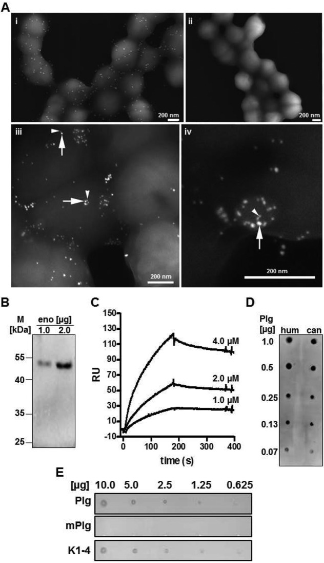
FIG 2.
Enolase of S. canis as surface-displayed plasminogen binding protein (A) (i) FESEM visualization of enolase on the surface of S. canis G2 using antienolase antibodies and protein A-gold (15 nm; immune gold labeling). (ii) S. canis G2 treated with preimmune serum and protein A-gold (15 nm) exhibits no labeling. (iii and iv) Visualization of enolase using immune gold labeling (10 nm) (arrows) and visualization of plasminogen binding to enolase by plasminogen-gold (15 nm) (arrowheads). (B) Western blot analysis of plasminogen binding to purified S. canis enolase (eno) after separation of 1.0 μg and 2.0 μg protein via SDS-PAGE. Plasminogen binding was detected with plasminogen-specific antibodies and horseradish peroxidase-conjugated secondary antibodies. Visualization of binding signals was performed using enhanced chemiluminescence detection. (C) Determination of dissociation constant describing plasminogen binding to S. canis enolase via surface plasmon resonance analyses using 4.0 μM, 2.0 μM, and 1.0 μM concentrations of enolase as an analyte (KD of 8 × 10⁻⁷ M). (D) Detection of enolase binding to human (hum) and canine (can) plasminogen using the dot spot technique after immobilization of 0.07 μg, 0.13 μg, 0.25 μg, 0.5 μg, and 1.0 μg of plasminogen isotypes onto a nitrocellulose membrane. Signal detection was performed with enolase-specific antibodies followed by peroxidase-conjugated secondary antibodies and enhanced chemiluminescence detection. (E) Dot spot overlay analysis was performed after immobilization of recombinant S. canis enolase in amounts of 10.0 μg, 5.0 μg, 2.5 μg, 1.25 μg, and 0.625 μg onto a nitrocellulose membrane. Human plasminogen (Plg), miniplasminogen (mPlg), representing kringle domain 5 and the enzymatic domain, and kringle domains 1 to 4 (K1-4), representing the first four kringle domains at the plasminogen N terminus, were used for protein overlay. Binding was detected with polyclonal plasminogen-specific antibodies and peroxidase-conjugated secondary antibodies followed by enhanced chemiluminescence detection.

