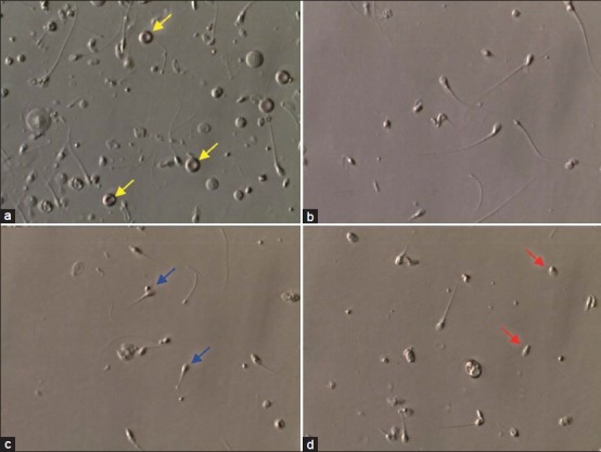
Figure 2.

Photomicrograph showing the overall pattern of epididymal specimens obtained by percutaneous epididymal sperm aspiration (PESA). (a) Raw specimen showing contamination with debris and red blood cells (yellow arrows). (b) Processed specimen after dilution and washing with erythrocyte lysing buffer solution. (c) and (d) Raw specimens obtained from the epididymis cauda. Aspirates form the cauda are often rich in senescent spermatozoa (blue and red arrows). Photographs obtained from the inverted microscope equipped with Hoffman modulation phase contrast (Nikon Diaphot 300) under ×400 magnification
