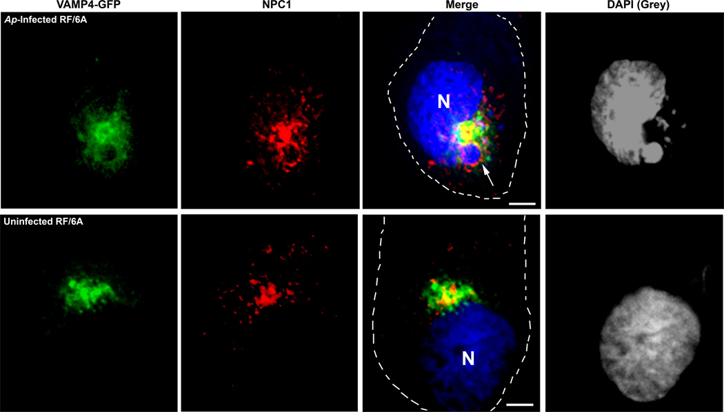
Fig. 7. VAMP4 and NPC1 are co-localized to A. phagocytophilum inclusions.
RF/6A cells were transfected with VAMP4-GFP plasmid and inoculated with host cell free A. phagocytophilum at 4h post transfection. At day 2 pi, the cells were fixed and stained with anti-NPC1 antibody and DAPI. VAMP4-GFP (green), NPC1 (red) and DAPI (blue) were visualized by DeltaVision deconvolution microscopy. A. phagocytophilum inclusions were visualized by DAPI staining (blue); the blue/DAPI staining was transformed to gray pseudocolor for clearer viewing of bacterial inclusions. The experiment shown is representative of three independent experiments. Bar, 5 µm. Ap, A. phagocytophilum. N, nucleus.

