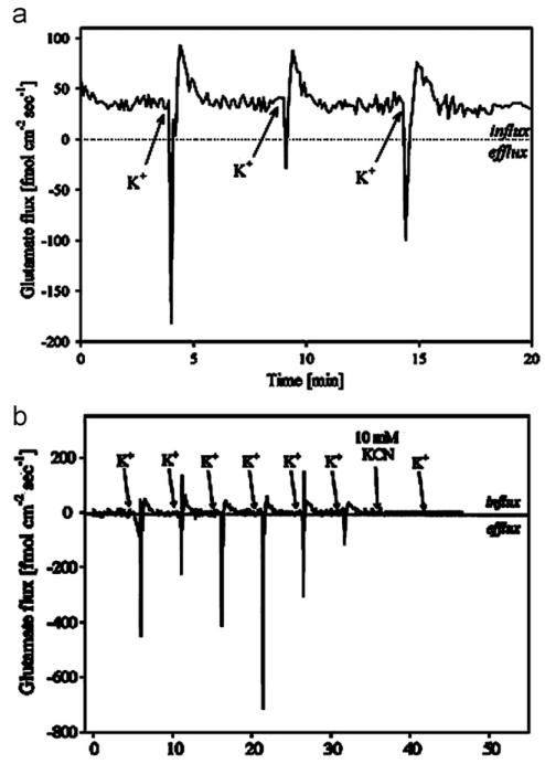
Fig. 4.
(a) Flux recorded 1 μM above the cells during three successive potassium stimulations with 150 μL of Locke’s buffer containing 53 mM KCl. Increase in Glu concentration and subsequent decrease in concentration were observed following stimulation. Phase sensitive detection provided signal noise filtration required to quantify biophysical glutamate efflux and influx. (b) Potassium stimulation of neural cells following 30 min of exposure to 100 μM threo-β-benzyloxyaspartate (TBOA). (Reprinted with permission from (McLamore et al. 2010b)).

