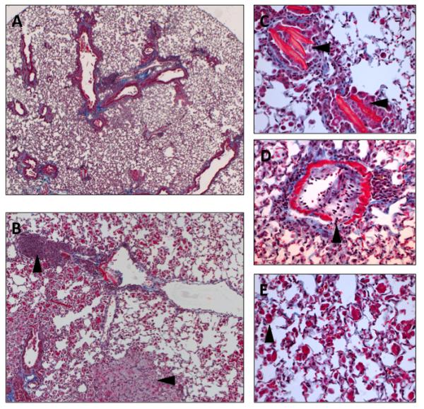
Figure 7. Obliterative bronchiolitis and interstitial pneumonia in C57L/J mice 26 weeks following 7.5 Gy to the whole thorax.

A) Masson’s Trichrome stain of lung tissue 26 weeks after 7.5 Gy whole thorax irradiation in C57L/J mice (5x magnification). B) Higher magnification (10x magnification) shows alveolar inflammation with foamy-macrophages (−>)and perivascular lymphocytic cuffing (*). C) Fine, eosinophillic needle like structures within the terminal bronchioles. D) Breakdown of the vessel wall, inflammation, and fibroproliferation. E) Giant, eosinophilic alveolar macrophages characteristic of alveolar inflammation. C-E, 40x magnification.
