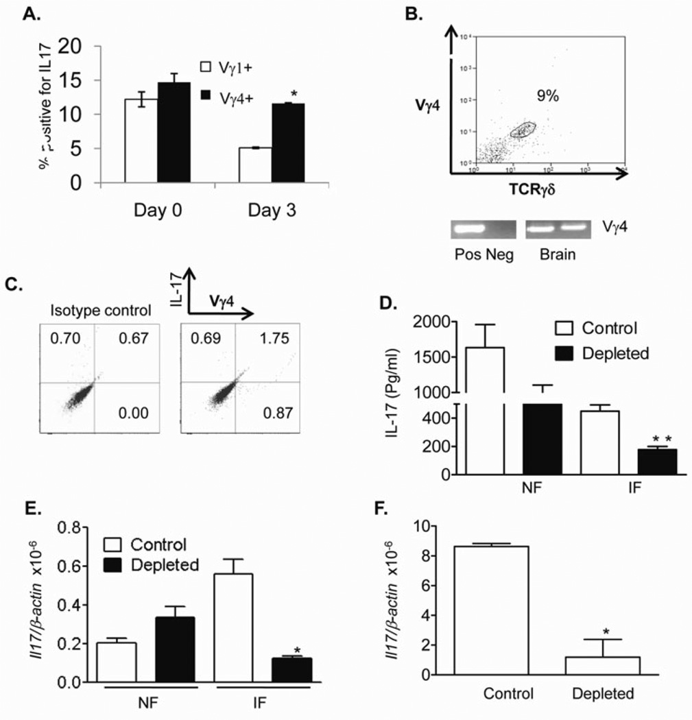
Figure 2. Vγ4+ T cells produced IL-17 during WNV infection.
A. Splenic T cells of naïve (day 0) or day 3-infected mice were cultured ex vivo with PMA plus ionomycin and stained for Vγ1 or Vγ4, and IL-17 and gated on each γδ T cell subset for analysis of the percentage of Vγ1+ IL-17+ or Vγ4+ IL-17+. n = 3–4. B. Vγ4+ T-cells were detected in WNV-infected mouse brains (day 7) by flow cytometry (top panel) and PCR analysis (bottom panel). Top panel: Cells were gated on CD3+CD45+ leukocytes. Bottom panel: Brain cDNA samples from two WNV-infected mice were used. Spleen cDNA was used as a positive control (Pos). Negative control (Neg): non-infected mouse brain. C. Brain leukocytes of day 7-infected mice were cultured ex vivo with PMA plus ionomycin and stained for Vγ4 and IL-17 and gated on total leukocytes for analysis of the percentage of Vγ4+ IL-17+. D. Supernatant of splenic T cells of non-infected (NF) or WNV-infected mice (IF) were treated with PMA plus ionomycin for 24 h and measured for IL-17 production, n = 7. E-F. Vγ4+-cell-depleted mice had a reduced IL17 production in blood (E) at day 3 and in brain (F) at day 7 post-infection, as determined by Q-PCR. n = 3–4. *P < 0.05 or **P < 0.01 for control vs. Vγ4+-T-cell-depleted mice.

