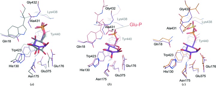
Figure 3.
Superposition of the phosphate- and glycon binding sites. The active sites of SmBgl–BG6 (purple) with (a) 6-P-β-galactosidase from L. lactis in complex with 6-P-β-galactose (gray; PDB entry 4pbg), (b) β-glucosidase from an uncultured bacterium in complex with β-glucose (pink; PDB entry 3fj0) and (c) 6-P-β-glucosidase A from E. coli in complex with a sulfate ion (PDB entry 2xhy) are shown.

