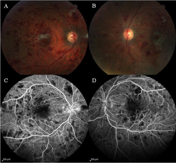
Figure 2.

Fundus examination at 6 weeks follow-up. Color fundus photograph of the right (A) and left eye (B) showing mild decrease in the intraretinal hemorrhages, cotton wool spots, and perifoveal whitening. Fluorescein angiography is showing the blocked areas corresponding to the hemorrhages (C, D) in late phase with mild decrease in the enlarged foveal avascular zone in both eyes compared with Figure 1C,D.
