Figure 2.

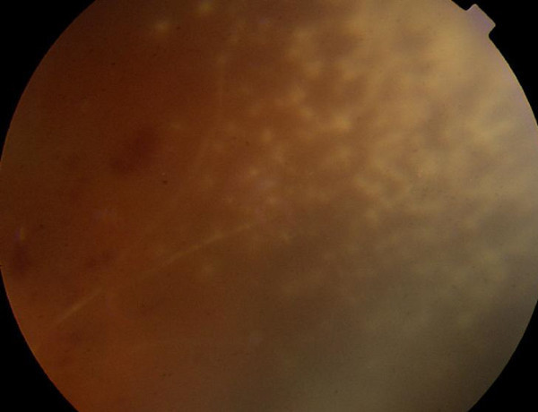
Case 1, superonasal retina, right eye. There are nummular patches of a granular necrotizing retinitis. A sclerotic arteriole enters the area. There are scattered intraretinal hemorrhages proximal to the optic nerve head.

Case 1, superonasal retina, right eye. There are nummular patches of a granular necrotizing retinitis. A sclerotic arteriole enters the area. There are scattered intraretinal hemorrhages proximal to the optic nerve head.