
Figure 3.
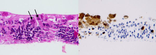
Case 1, retinal biopsy. Left frame: hematoxylin and eosin stain of retinal biopsy specimen showing cytomegalovirus inclusion bodies (arrows), ×100. Right frame: immunohistochemical stain with anti-cytomegalovirus antibody and avidin-biotin-peroxidase complex, 100X. Brown stain indicates retinal cells positive for cytomegalovirus antigens. Figures courtesy of Sander Dubovy, MD.