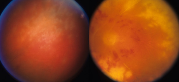
Figure 6.

Case 3, fundus, left eye. Left frame: a single small focus of a granular necrotizing retinitis was present in the superotemporal retina at the time of central retinal artery occlusion. Right frame: typical CMV retinitis developed several weeks later after discontinuation of systemic anti-viral therapy with oral ganciclovir.
