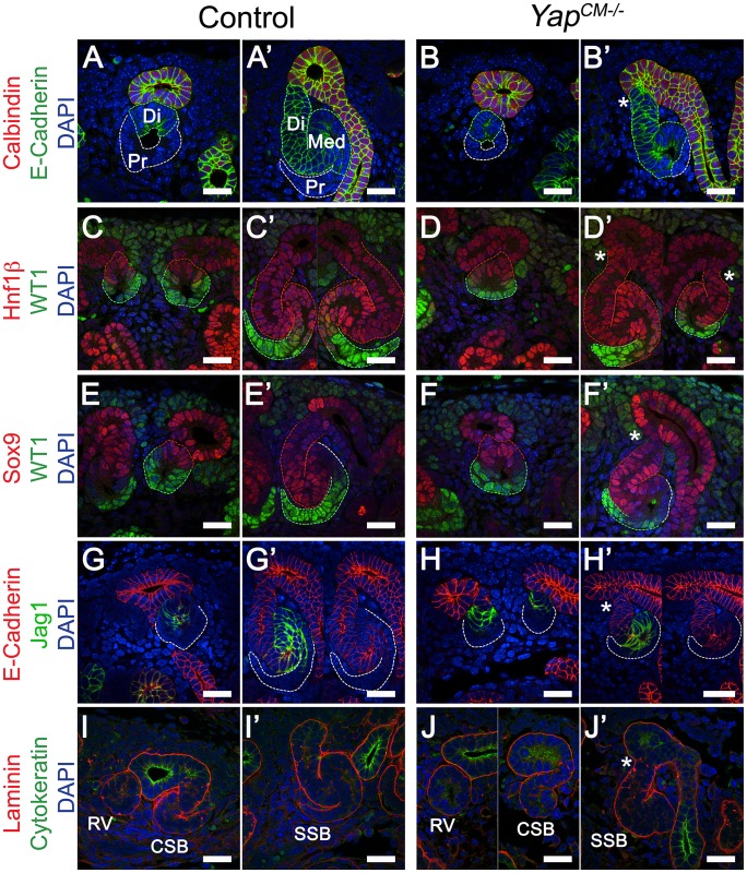
Figure 4. Characterization of segmentation in Yap mutant nephrons.
(A–B′) Double staining for E-cadherin and Calbindin in RV and SSB. Co-staining for Hnf1ß/WT1 (C–D′) and Sox9/WT1 (E–F′) reveals normal segmentation of the RV with both proximal and distal segments. Similarly, SSB show normal segmentation. Note the reduced size of the proximal domain in Yap-null SSB (compare WT1 positive segment in Yap mutants (D′, F′) to controls (C′, E′). This is also apparent in B′ and J′. (G–H′) Immunofluorescence for E-cadherin and Jag1 reveals no change in specification of the distal RV and the medial segment of the SSB in both genotypes. Note the aberrant morphology (asterisk) of the site where the connection occurred between the SSB and the UE (B′,D′,F′,H′ and J′). (I–J′) Immunofluorescence using antibodies to Cytokeratin (UE) and Laminin (BM) shows that fusion occurred before the comma-shaped stages. All staining performed at E15.5. CSB: comma-shaped body; RV: renal vesicle; SSB: S-shaped body. Scale bars represent 25 µm. DAPI was used to counterstain nuclei.

