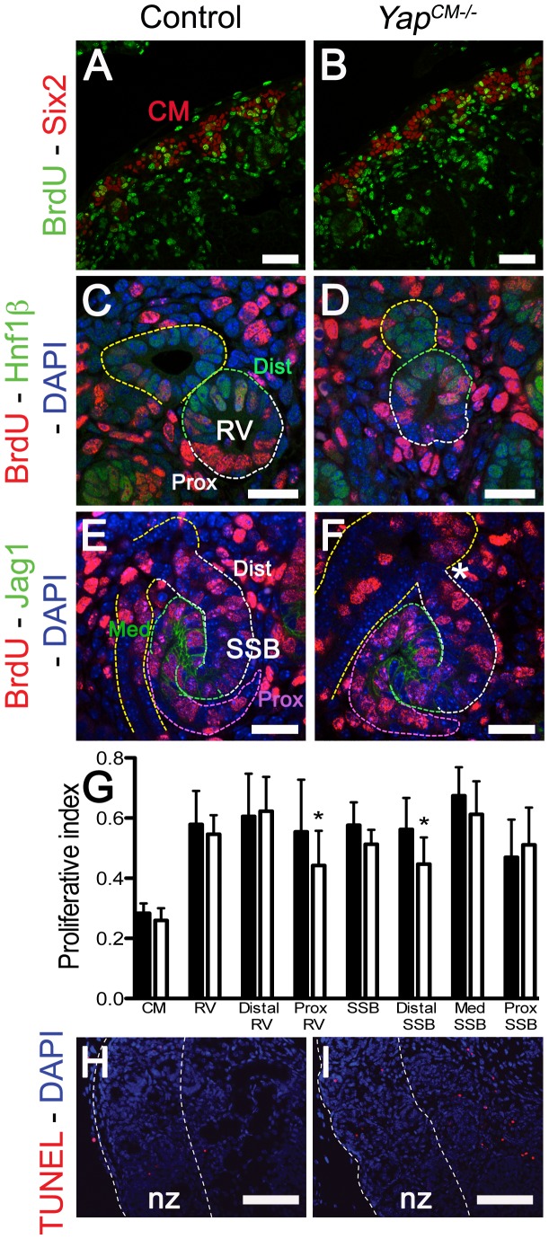
Figure 5. No major change in apoptosis or proliferation in Yap mutant kidneys.
Confocal images of BrdU incorporation in condensing mesenchymal cells (A,B), renal vesicle (C,D) and SSB (E,F) at E15.5. (A,B) Co-staining with Six2 antibody was used to co-labeled cap mesenchyme cells. (C,D) Co-staining with Hnf1ß antibody was used to distinguish the distal (Dist) from the proximal (Prox) segment of the RV. (E,F) Jag1 antibody was used to identify the distal (Dist), medial (Med) and proximal (Prox) segments of the SSB. (G) Quantification of the proliferation index in controls (black columns) and Yap mutants (white columns) throughout nephrogenesis. Prox RV*: p = 0.0319; Distal SSB*: p = 0.0353. (H,I) TUNEL assay at E18.5 reveals no change in apoptosis in mutant nephrogenic zone (nz). There is often an increase in apoptosis in the later developing inner cortex in the Yap mutants. Scale bars represent 50 µm (A,B), 25 µm (C,F), 100 µm (H,I).

