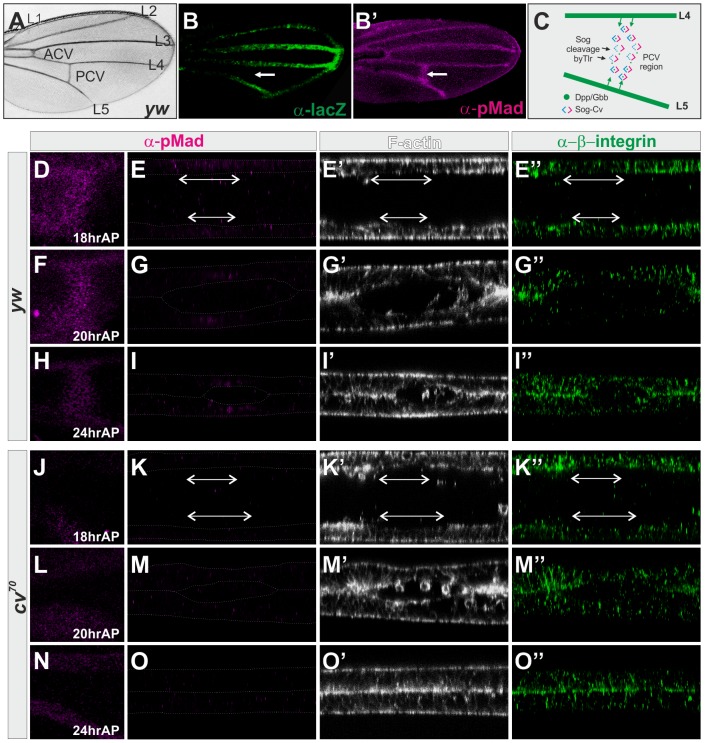
Figure 1. BMP signaling is required for PCV morphogenesis.
(A) Wild-type yw adult wing. (B, B′) lacZ (B) and pMad (B′) staining of dppshv-lacZ at 24 hr AP. The PCV position is indicated by arrows. (C) A current model of Sog-Cv-mediated directional BMP transport from the LVs into the PCV. (D—I) Wild-type yw pupal wing. pMad (D, E, F, G, H, I), F-actin (E′, G′, I′), and ß-integrin (E″, G″, I″) staining at 18 hr AP (D, E), 20 hr AP (F, G), and 24 hr AP (H, I). (J—O) cv70 pupal wing. pMad (J, K, L, M, N, O), F-actin (K′, M′, O′), and ß-integrin (K″, M″, O″) staining at 18 hr AP (J, K), 20 hr AP (L, M), and 24 hr AP (N, O). (D, F, H, J, L, N) Dorsal view of the PCV region. (E, G, I, K, M, O) Optical cross-sections of the PCV region. Prospective PCV positions are indicated by double-headed arrows at 18 hr AP (E–E″, K–K″).

