Figure 2.

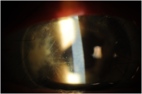
Slit lamp photograph two weeks after initial presentation. This shows the resolution of the epithelial defect. The intralamellar branches remain.

Slit lamp photograph two weeks after initial presentation. This shows the resolution of the epithelial defect. The intralamellar branches remain.