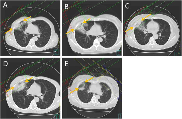
Figure 1.

Fusion of CT images at the time of diagnosis of OP and treatment planning images (A = patient 1, B = patient 2, C = patient 3, D = patient 4, E = patient 5). There were free regions between the radiation lesions and the radiation-induced OP (arrow).
